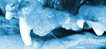
Veterinary Dental Center

Oral Mass 101

(Jan/Feb 2007)

Mandibular Fracture Management in AnimalsOral tumors frequently go unnoticed in pets until they reach an advanced stage. Unfortunately, the gross appearance (smooth vs. ulcerated vs. pigmented tissue) of a mass does not indicate the tumor type, rather a biopsy is the mainstay for diagnosis and decision making. Therefore, a systematic approach in the diagnosis of an oral mass in our feline and canine patients is important in terms of treatment and prognosis.

The initial evaluation of an oral mass includes an evaluation of the location and size of the mass, blood and urine profile to rule out systemic disease, evaluation of regional lymph nodes and thoracic radiographs to detect any signs of distant metastasis.

Next, an oral examination under anesthesia and intra-oral radiographs are performed. Depending on the size, location and evidence of bone involvement, an incisional or excisional biopsy is performed. Biopsy specimens are taken from the center of the mass and should include bone if the lesion is present on radiographs. Fine needle aspirates of the mandibular lymph node or in select cases an excisional biopsy of the ipsilateral parotid, retropharyngeal, and mandibular lymph nodes are used to assess metastasis.

The tumor size, tumor type (identifies it’s biologic behavior) and results of staging will direct the treatment strategy. For example, a squamous cell carcinoma of the gingiva offers a better prognosis than one of the tongue or tonsillar region. Moreover, a malignant melanoma metastasizes early, a squamous cell carcinoma metastasizes late, and an acanthomatous epulis is benign and only locally invasive. The client will be counseled on the prognosis, post procedure appearance and function prior to treatment.

Surgical excision is the preferred treatment in the majority of cases. For benign masses without bone involvement, a marginal excision is adequate (1-5 mm of normal tissue). Malignant masses require surgical treatment by the means of mandibulectomy or maxillectomy (1-2 cm of normal tissue).

Common Oral Masses

Benign - Fibromatous epulis

  • Ossifying Epulis
  • Acanthomatous Epulis
  • Cysts - Dentigerous cyst
  • Gingival cyst
  • Eruption cyst
  • Odontoma
  • Ameloblastoma
  • Malignant - Malignant melanoma
  • Squamous cell carcinoma
  • Fibrosarcoma
  • Osteosarcoma
  • Plasma cell tumor
  • Hemangiosarcoma Inflammation - Gingival Hyperplasia
  • Fibromucosal Poly
  •