Enamel Hypoplasia
When the enamel (ie. outer layer of a tooth) fails to develop properly, your pet’s teeth may be at risk for infection or fracture. This condition is known as enamel hypoplasia.
What you need to know…
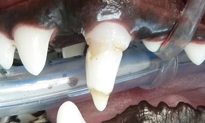
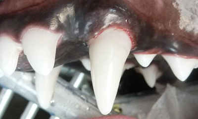
- Teeth will appear rough, pitted and stained yellow.
- Your pet may experience heightened sensitivity.
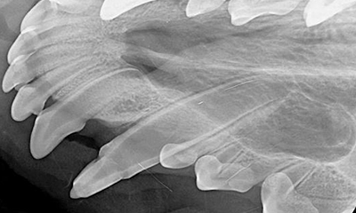
- Teeth will be more prone to tartar accumulation, gum disease, infection and may have abnormal root development.
How Enamel Hypoplasia may look:
Possible Therapy / Treatment:
Depending on the severity of the enamel defect and the functional importance of the affected tooth, extraction or crown placement are possible treatments.
Schedule an appointment to have your pet evaluated by clicking on the “Get In Touch with Us” button.
Learn More
Enamel hypoplasia refers to a condition in which the outer layer of the tooth (enamel) fails to develop properly. These teeth will appear rough, pitted and stained yellow. These pets can have sensitivity, are more prone to tartar accumulation, gum disease, infection and may have abnormal root development.
These pets need dental x-rays, tooth debridement and either: sealant, composite restoration, extraction or crown placement. The treatment decisions are based on the severity of the enamel defect, the functional importance of the tooth, and your pet’s lifestyle.