3D Imaging - Cone Beam CT
KEY FACTS
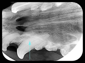
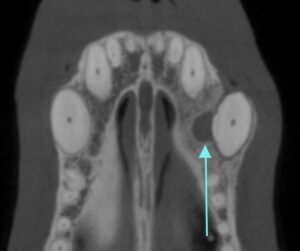
- Cone beam CT is a rapid, non-invasive scan that allows highly accurate 2D and 3D imaging of the teeth, bone and skull anatomy
- Allows us to identified many hidden conditions not visible on dental radiographs (x-rays)
- Essential imaging tool for patients with: tooth fractures, bone loss, missing teeth, tooth crowding, facial swelling, nasal discharge, evaluation of an oral mass/growth, TMJ disease and facial trauma (jaw fracture)
QUICK LOOK AT 3d imaging


LEARN MORE
The Veterinary Dental Center utilizes CBCT imaging for the majority of their patients. It provides the highest quality imaging which can be evaluated in multiple planes, with unlimited contrast and assessed using 3D maxillofacial reconstruction. It routinely identifies many oral conditions that may not be visible on dental radiograph (x-rays) as well as reduces time under anesthesia.
Our CBCT unit, VetCAT, is an FDA approved imaging tool that is mobile, allowing us to perform a scan in 40 seconds tableside without moving the patient. This optimal workflow minimizes anesthetic time, allows the doctor to evaluate the scan/images in the treatment room and efficiently communicate to the pet’s dental team the diagnosis and treatment plan for the pet.