Dental X-ray
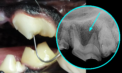
Dental x-ray is an essential tool in the diagnosis and treatment planning of oral conditions in your pet as 60% of the tooth lies under the gums.
KEY FACTS
- 60% of the tooth lies under the gums and must be evaluated with dental x-rays
- x-rays allow assessment of the tooth, root, bone as well as adjacent structures (teeth, nasal cavity, sinus, blood vessels/nerves)
- Digital dental x-ray system is used for superior image quality and it reduces radiation exposure to the patient.
QUICK LOOK AT DENTAL x-RAY
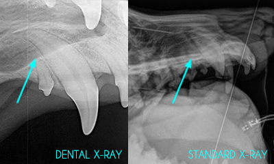
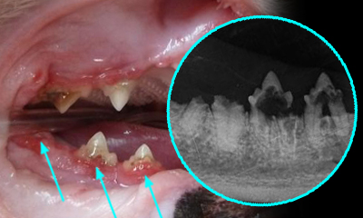
The canine tooth below illustrates how much more detail can be obtained with a dental x-ray vs. standard x-ray system.
LEARN MORE
Dogs have 42 teeth and cats have 30 teeth. Usually, 8 to 15 x-ray images are obtained in order to effectively access oral condition. x-rays will allow us to possibly discover:
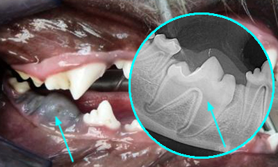
- Extra root or an abscess that has extended to an adjacent/normal looking tooth.
- Unerupted or impacted teeth, which can lead to a bone cyst.
- Teeth that must be extracted need to be evaluated for root fractures or root ankylosis (fusion to the bone). Also, post-extraction x-rays are the standard of care in dentistry.
- Cats are prone to “cavities” or resorptive lesions. Some lesions are located under the gums and are only visible on x-rays. Treatment is based on the evaluation of root structure, where intact roots must be extracted, while resorbing roots can be retained.
- Oral growths (cyst, infection or tumor) require x-ray assessment for treatment or biopsy.